PART 1.4 - CORE CONCEPTS
How are x-rays used in archaeology?
X-rays are used in a wide manner of methods within archaeological research. For the purposes of clarity when I refer to 'X-rays' I really mean the production of radiographs through the application of radiography. An assortment of different technologies use X-rays as shall be shown in Part 4 Paleoradiography in research.
A short history lesson is required to address how X-rays are used in archaeology.
The discovery of X-rays by Wilhelm Röntgen in 1895 led to incredible developments in medicine and patient care. The utility of X-rays to archaeology did not go unnoticed, and in 1896 Carl Koenig, a lecturer in physics at Frankfurt (Germany), used the new radiation to image the knees of an ancient Egyptian child mummy. Many other investigators followed suit, imaging Egyptian and Peruvian mummies to discover what lay beneath without having to conduct an autopsy (the infamous unwrapping ceremonies popular with the Victorians (!)).
It is difficult to comprehend the way X-rays revolutionised medical and scientific progress. It was truly a game-changer. Modern technologies such as computed tomography (CT) provide three-dimensional imaging with huge advantages over radiography. It is little wonder that current paleoradiology research is dominated by CT.
Despite the obvious advantages of CT within archaeological research the use of radiography persists, and for good reason. Aside from being relatively cheaper, digital radiography has lower logistical burdens along with lower training requirements. The video below (!) provides a brief explanation for the difference between CT and radiography.

When is radiography used in archaeology?
This depends on what has survived the test of time and has been excavated or recovered from antiquity for scientific inquiry. In my opinion, there are three types of materials that may undergo radiography:
Bones (human or animal)
Metalwork (weapons, tools, vessels or jewellery)
Ceramics (commonly recovered, uncommonly investigated by paleoradiography)
I have made a glaring omission - the imaging of mummies. Mummies can be loosely termed as human remains with preserved soft tissues. They are often prized possessions, rare and of significant scientific value. However, their rarity makes them outside of routine imaging within archaeology. They also tend to suffer from the 'gee-whizz, I wonder what we can see if we X-ray it' syndrome. The lack of scientific investigation is often apparent in early literature, where mummies underwent CT scans just to see what was inside rather than to answer specific hypotheses. I have also omitted other perishable materials such as wood and leather due to the relative rarity of survival in the archaeological record, although reported examples exist (especially within glacial archaeology).
The use of radiography in archaeology according to Historic England:
From my own research I have encountered one document produced by Historic England that explicitly explains the use of radiography in archaeology - Guidelines on the X-radiography of archaeological metalwork (2006).
The use of radiography has also been mentioned in many guidance documents by Historic England:
Waterlogged organic artefacts (2018)
Animal bones and archaeology: Recovery to archive (2019)
Archaeological evidence for glassworking (2018)
Archaeometallurgy: Guidelines for best practice (2015)
Geoarchaeology: Using earth sciences to understand the archaeological record (2015)
If you are aware of other guidance documents for the use of radiography in archaeology can you please let me know by contacting me? I am particularly interested in how radiography is used in other countries (outside of the United Kingdom). I will add links for future participants.
Books about paleoradiography (and paleoimaging in general):
There are some academic textbooks available which explore the use of radiography in archaeology. Some may be available through your university or public library.
Paleoimaging: Field Applications for cultural remains and artifacts
By Ronald G. Beckett & Gerald J. Conlogue
2010 CRC Press
PaleoRadiology: Imaging mummies and fossils
By Rethy K. Chhem & Don R. Brothwell
2008 Springer
Advances in paleoimaging: Applications for paleoanthropology, bioarchaeology, forensics, and cultural artifacts
By Gerald J. Conlogue & Ronald G. Beckett
2020 CRC Press
Case studies for advances in paleoimaging and other non-clinical applications
By Ronald G. Beckett, Gerald J. Conlogue & Andrew Nelson
2020 CRC Press
What can we learn from radiography in archaeology?
The information below outlines what we can learn using radiography upon materials typically recovered within archaeology. Many of these issues will be further explored in later parts of this course, alongside published case studies.
I've used my experience and research to inform what is shown below. I admit it is not an exhaustive list of research possibilities. There are other reasons for using radiography in archaeology, but I feel these are the main reasons.
In theory, the use of X-rays may destroy or damage ancient DNA (as shown by this article) but the levels of radiation involved in radiography are comparatively very low.

Bone:
This is not restricted to human bones, it may include animal bones too. Additionally, it may include the use of bone as a material used in manufacturing rather than just funerary or cooking remains. I have provided links to literature for examples of radiographs and their scientific investigation. Further examples can be found in Part 4.3.
Pathologies - Specifically, the identification of cancers, bone infections and genetic abnormalities suspected by visual inspection and confirmed through radiographic imaging. Characterisation of pathologies, such as size, shape and extent of spread, are all reasons for X-rays. Lastly, undiagnosed conditions that cannot be identified through visual inspection alone often benefit from imaging for clues. This article provides an osteobiography of a 14th century Chilean population. The researchers use radiography to help identification of pathologies.
Trauma - Again, may already be noticeable through visual inspection but may benefit from radiographic characterisation. Sometimes subtle fractures (new or healed) may be seen on X-rays. Lastly, foreign bodies such as weapon fragments (metal, stone, possibly wood) can be identified in situ. An article by Dittmar et al (2020) presents medieval injuries which includes radiographs.
Stress - This relates to biological stress demonstrated upon bone development or maintenance. For example, skeletons may exhibit osteoporosis (loss of bone mineral density), interruptions to bone growth during adolescence (as Harris lines) or failure to develop due to pathological or nutritional detriments. Nowakowski (2018) provides an example of radiography being used for Harris line investigation of an ancient bear.
Age - A common tool within forensic imaging, radiography of the teeth can provide an accurate estimation of age. Although the eruption of teeth stop in your late 20's they accrue damage through wear and tear. Radiography may also demonstrate such attrition, but its use as an age estimation is not as accurate as different populations experienced different levels of tooth decay/loss. For example, ancient populations such as the Egyptians used sand (or other coarse material) during milling of flour. The sand therefore made its way into bread and caused damage to teeth. A review on dental aging can be found here. An archaeological example of dental wear of Viking-age Icelanders can be found here, although they do not use radiography.


Metalwork:
Radiography has been used on a wide variety of metal items ranging from weapons to tools to precious items. Metal items often suffer from deterioration through corrosion or rusting within the earth, leaving either very fragile and fragmented remains or a conglomerate mass of soil and oxides. Non-ferrous metals are the exception, with gold and silver both surviving incredibly well.
Identification - Simply put, to identify what the item is (see below). This has a two-fold purpose: firstly as a triage method for determining whether the item should be catalogued, conserved and curated. Common items such as nails may not be kept but items of status or worth may warrant the expense of cleaning, investigation and possibly restoration. The second purpose of identification would be to date the period of occupation or characterise the land use (e.g. rural or urban).
Conservation - Going hand-in-hand with identification is the use of X-rays with conservation efforts. Having an image before and after cleaning provides evidential proof of the item status. If it was broken before, it was going to be broken after the soil/oxides were removed! On a less cynical point of view, X-rays may aid the conservator by identifying areas of structural weakness. It is also much easier to prioritise efforts for item extraction or salvage if you know where to look (as may be the case with soil-blocks). Check out this open access article demonstrating the use of CT for a soil block containing a metal archaeological artefact. The authors initially use radiography to identify and localise the metalwork.
Construction/production methods - Radiography may also elucidate clues as to the construction or production methods of metal items. Is it one solid piece or two? Are there rivets or screws? Are the components of different density? Knowledge of the production methods of the time period would be key to deciphering the X-rays.


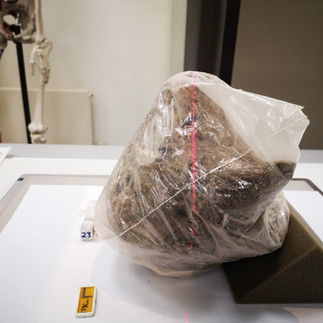
This collection of corroded metals showed several iron lumps (of unknown identity) and a solitary ring. Saxon Kent.
Ceramics:
Admittedly, not frequently investigated using radiography, but one of the most commonly found items during archaeological excavations in England. Ceramics may relate to everyday items such as pots, vases and various storage vessels. Funerary items could also be classed as ceramics, such as urns (see below). Rarely, there may be decorative items such as statues or inscribed/carved clay tablets. At a stretch, you may also consider carved rock. This article looks at archaeological pottery with industrial CT, providing a general overview of the process. The article by Agnieszka Mączyńska (2021) investigates Egyptian Predynastic pottery production with radiography.
Contents - Funerary urns may contain bones and metalwork. An X-ray helps to identify these prior to excavation. Alternatively, storage items may also contain stashes of coin or other items of value.
Conservation - As with metalwork, radiography has been used to image ceramics for the purpose of conservation. Museums may use X-rays to check the stability of items in their holdings to assess for damage or urgent repairs.
Construction / production methods - Again, radiography may provide clues to the construction and/or production methods of ceramics. For example, clay with large particles are radiographically different from clay with small particles. The same may also be said for ceramics made by hand-moulding when compared to those made on the potter's wheel.


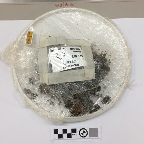
A Roman funeral urn showing irregular densities suggestive of bones. No metalwork could be found.
Reading task
Read the short article 'The earliest documented applications of X-rays to examination of mummified remains and archaeological materials' by Fiori and Nunzi (1995).
The article can be found using this link. I find it easier to download the PDF version to read the article.
This article illustrates the emergence of X-rays and how they were quickly adopted within archaeology.
Estimated reading time: 10 minutes.
